Welcome to our comprehensive guide comparing cephalomedullary nails and intramedullary nails for the treatment of femoral fractures. As a leading resource in orthopedic surgery, we are here to provide you with valuable insights into these two surgical techniques.
Fractures of the femur, particularly intertrochanteric fractures, require careful consideration and an effective treatment approach. In this article, we will explore the nuances of cephalomedullary nails and intramedullary nails, including their indications, surgical techniques, outcomes, and potential complications.
Key Takeaways:
- Cephalomedullary nails and intramedullary nails are two common surgical methods used to treat femoral fractures.
- Each type of nail has specific indications based on the type and location of the fracture.
- The surgical technique for cephalomedullary nails involves accessing the femur through the hip joint, while intramedullary nails are inserted directly into the medullary canal of the femur.
- Potential outcomes of both procedures include improved fracture stability and enhanced healing.
- Complications, although rare, can include infection, nail breakage, and nonunion of the fracture.
Now, let’s dive into a detailed exploration of cephalomedullary nails and intramedullary nails, starting with an in-depth look at cephalomedullary nails in the next section. Stay with us to gain a comprehensive understanding of these orthopedic surgical techniques and make informed decisions for your patients.
#cephalomedullarynail #intramedullarynail #femoralfractures #orthopedicsurgery #surgicaltechnique #outcomes #complications
Understanding Cephalomedullary Nails
In this section, we will focus on cephalomedullary nails, a type of implant commonly used in the treatment of femoral fractures. These specialized nails offer several advantages over other types of implants, making them a popular choice among orthopedic surgeons.
Indications:
Cephalomedullary nails are primarily used in the management of fractures involving the proximal femur, including intertrochanteric and subtrochanteric fractures. These implants are designed to provide stability and promote early weight-bearing, enabling quicker recovery and optimal functional outcomes for patients.
Surgical Technique:
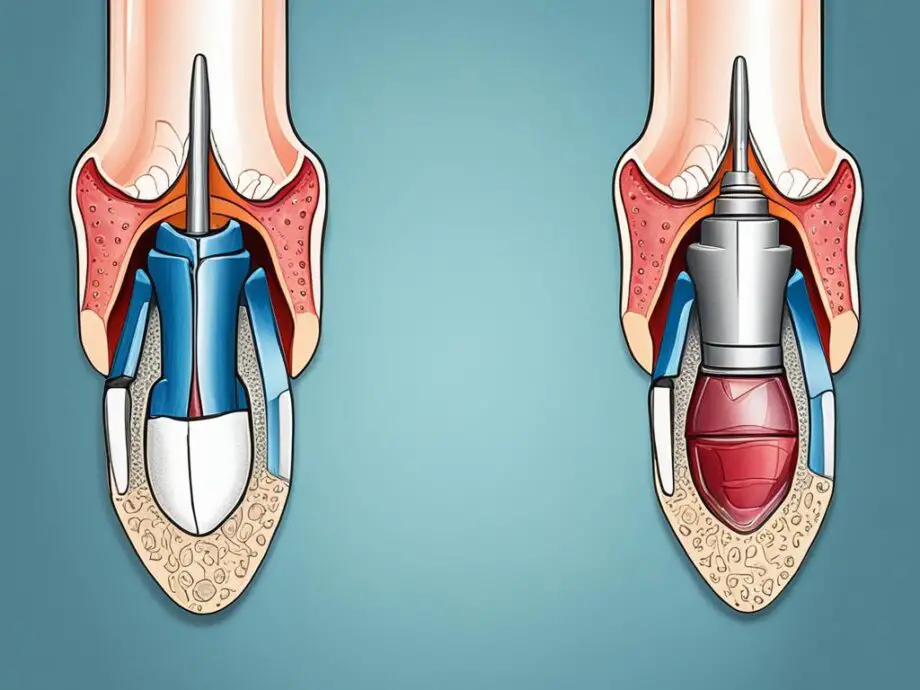
The placement of a cephalomedullary nail involves a minimally invasive surgical technique. A small incision is made and the nail is inserted into the medullary canal of the femur, extending from the femoral head to the distal shaft. The nail is secured with screws at both ends, providing stable fixation.
Outcomes and Complications:
Cephalomedullary nails have shown favorable outcomes in the treatment of femoral fractures, with high union rates and good functional outcomes reported in numerous clinical studies. However, as with any surgical procedure, complications can occur. These may include infection, implant failure, nonunion, malunion, and intraoperative fractures. Close postoperative monitoring and appropriate management of complications are essential for achieving successful outcomes.

Overall, cephalomedullary nails offer orthopedic surgeons a reliable and effective option for the treatment of femoral fractures. Their specific indications, minimally invasive surgical technique, and potential outcomes and complications make them a valuable tool in the management of these challenging injuries.
Intramedullary Nails: A Comprehensive Overview
When it comes to the treatment of femoral fractures, intramedullary nails have been widely used and proven to be an effective surgical intervention. These nails are inserted into the medullary canal of the femur, allowing for stability and alignment of the fractured bone.
Indications
Intramedullary nails are commonly employed in the treatment of both femoral fractures and intertrochanteric fractures. These fractures are often the result of high-energy trauma, such as motor vehicle accidents or falls from a height. Intramedullary nails are particularly suitable for these fractures due to their ability to provide superior fixation and load sharing capabilities.
Surgical Technique
The placement of intramedullary nails requires a careful surgical technique. First, a small incision is made at the proximal end of the femur to gain access to the medullary canal. The canal is then reamed to accommodate the nail, and the appropriate-sized nail is inserted. Once the nail is in place, locking screws are placed to provide additional stability.
Outcomes
The use of intramedullary nails has shown favorable outcomes in the treatment of femoral fractures. These nails allow for early mobilization, leading to quicker recovery and reduced hospital stays. Additionally, they provide excellent rotational stability, promoting better functional outcomes for patients.
Complications
While intramedullary nails are generally safe and effective, they are not without potential complications. Infection, malalignment, nonunion, and implant failure are some of the complications that can occur. However, with proper patient selection and adherence to surgical techniques, the risk of complications can be minimized.

Overall, intramedullary nails play a crucial role in the management of femoral fractures and intertrochanteric fractures. Their specific indications, surgical technique, and potential outcomes and complications highlight the importance of careful consideration and expertise in their use.
Comparing Cephalomedullary Nails and Intramedullary Nails
When it comes to the treatment of femoral fractures, orthopedic surgeons have two main options: cephalomedullary nails and intramedullary nails. While both these implants are used to stabilize fractured femurs, they differ in several key aspects, including their indications, surgical technique, outcomes, and potential complications.
Indications
Cephalomedullary nails are primarily used for the treatment of intertrochanteric fractures, where the fracture line is located between the greater trochanter and the lesser trochanter. On the other hand, intramedullary nails are more commonly employed for diaphyseal fractures, which occur in the long shaft of the femur.
Surgical Technique
The surgical technique for inserting cephalomedullary nails involves making an incision near the greater trochanter, followed by inserting the nail into the intramedullary canal. This allows for stabilization of the femur and proper alignment of the fracture fragments.
Intramedullary nails, on the contrary, require the surgeon to make an incision near the knee joint. A guide wire is then inserted through the femur and used as a pathway to place the nail within the medullary canal.
Outcomes
The choice between cephalomedullary nails and intramedullary nails can significantly impact the outcomes for patients with femoral fractures. Studies have shown that both types of implants provide effective stabilization, allowing for early mobilization and promoting fracture healing. However, the choice of implant should be based on individual patient factors, fracture morphology, and the surgeon’s expertise.
Complications
Complications can occur with any surgical procedure, and the use of cephalomedullary nails and intramedullary nails is no exception. Potential complications include infection, nonunion (failure of the fracture to heal), malalignment, hardware failure, and avascular necrosis. However, with proper patient selection, surgical technique, and postoperative care, the incidence of these complications can be minimized.
By understanding the differences between cephalomedullary nails and intramedullary nails, orthopedic surgeons can make informed decisions to provide the most appropriate treatment for their patients with femoral fractures.

Next, we will summarize the key points discussed in this article regarding the comparison between cephalomedullary nails and intramedullary nails.
Conclusion
In summary, this article has explored the key differences between cephalomedullary nails and intramedullary nails for the treatment of femoral fractures. Both types of nails have their specific indications, surgical techniques, and potential outcomes and complications.
For cephalomedullary nails, they are commonly used for unstable intertrochanteric fractures and are inserted into the femoral head or neck. The surgical technique involves making an incision over the greater trochanter and placing the nail through the medullary canal. Potential outcomes include improved stability and fracture healing, but complications such as implant failure and cut-out may occur.
On the other hand, intramedullary nails are often employed for femoral shaft fractures and are inserted into the femoral canal. The surgical technique entails reaming the canal and inserting the nail through a small incision. Possible outcomes include good alignment and early mobilization, while complications such as infection and malalignment may arise.
Overall, the choice between cephalomedullary nails and intramedullary nails depends on the specific fracture pattern and patient characteristics. Surgeons must carefully consider the indications, surgical techniques, and potential outcomes and complications associated with each type of nail in order to make the best decision for their patients.
FAQ
What are the main differences between cephalomedullary nails and intramedullary nails?
Cephalomedullary nails and intramedullary nails differ in their design and approach. Cephalomedullary nails have a proximal anchoring screw that provides additional stability in femoral fractures, while intramedullary nails rely on distal locking screws. Furthermore, cephalomedullary nails are specifically designed for the treatment of intertrochanteric fractures, whereas intramedullary nails can be used for various types of femoral fractures.
What are the indications for cephalomedullary nail placement?
Cephalomedullary nails are primarily indicated for intertrochanteric hip fractures. These include pertrochanteric and subtrochanteric fractures, where the fracture line extends into the trochanteric area of the femur.
How is the surgical technique for cephalomedullary nail placement performed?
The surgical technique for cephalomedullary nail placement involves a lateral approach to the hip. After exposing the fracture site, the nail is inserted into the femoral canal and anchored with a proximal screw. Distal locking screws are then placed to provide stability. Fluoroscopic guidance is typically used throughout the procedure to ensure precise nail placement and screw fixation.
What are the potential outcomes and complications associated with cephalomedullary nail use?
Cephalomedullary nails have shown favorable outcomes in the treatment of intertrochanteric fractures, including improved functional outcomes and a high rate of fracture union. However, complications such as cut-out or breakage of the proximal screw, infection, nonunion, and implant-related complications can occur.
What are the indications for intramedullary nail placement?
Intramedullary nails are commonly indicated for femoral shaft fractures, both in the diaphysis and metaphysis. They offer stability and support for long bone fractures, facilitating early mobilization and reducing the risk of nonunion.
How is the surgical technique for intramedullary nail placement performed?
The surgical technique for intramedullary nail placement involves the insertion of a nail through the medullary canal of the femur. The nail is secured with locking screws at the proximal and distal ends of the fracture site. This technique provides stability and allows for biological fracture healing.
What are the potential outcomes and complications associated with intramedullary nail use?
Intramedullary nails have shown excellent outcomes in the treatment of femoral fractures, including high union rates and early mobilization. However, potential complications include infection, malunion, nonunion, implant failure, and knee pain.
How do cephalomedullary nails and intramedullary nails compare?
Cephalomedullary nails and intramedullary nails differ in their specific indications, surgical techniques, and potential outcomes and complications. Cephalomedullary nails are designed for intertrochanteric fractures, while intramedullary nails are more versatile and can be used for various femoral fractures. The choice of nail depends on the specific fracture type and patient factors.